Six Bodies That Betrayed Themselves
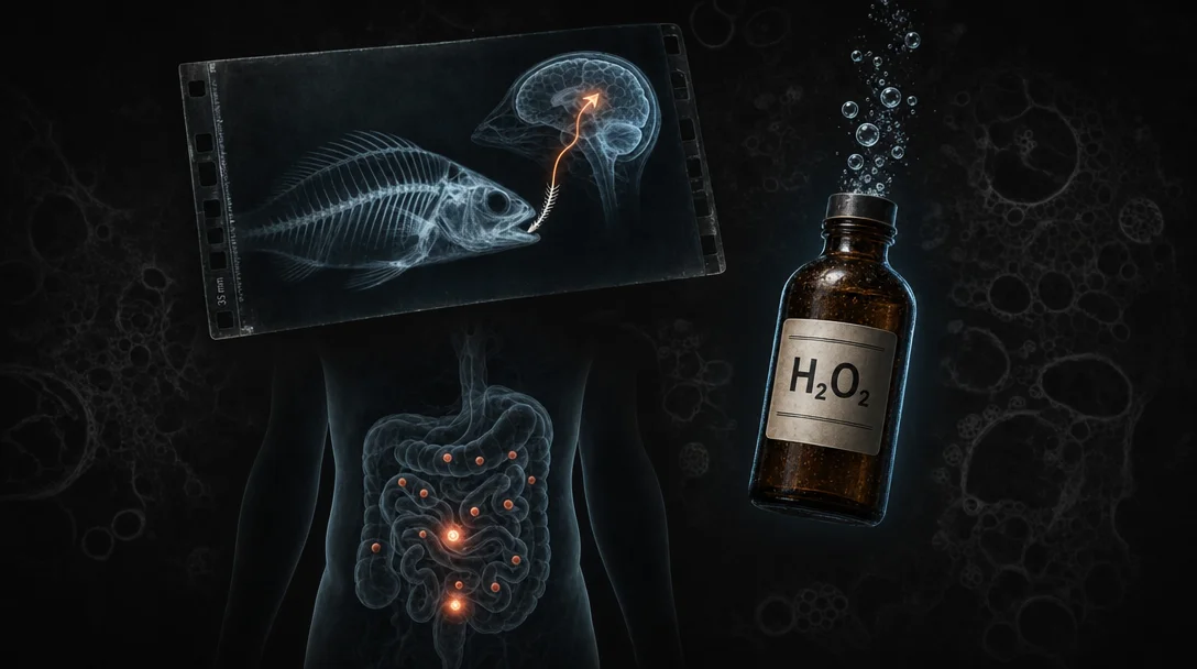
Six clinical case reports from BMJ Case Reports, June 1–8, 2026: a swallowed fish bone that fistulated into a pulmonary vein and sent air bubbles to the brain; hydrogen peroxide wound irrigation that stopped a man's heart on the table; thirteen silent jejunal tumors found only when the surgeon counted; the Leser-Trélat sign — a sudden eruption of hundreds of warty lesions that vanished when the cancer was treated; nivolumab+ipilimumab triggering Graves' disease and thyroid storm; and a woman whose shingles reactivation added a fourth simultaneous autoimmune condition.
Six cases from this week's journals — June 1 through 8, 2026 — where the body's own mechanisms did most of the damage. A bone opened a highway to the brain. A wound irrigant stopped a heart. A cancer signaled its presence through a sudden rash, then erased the evidence when it was treated. The mechanism in each case was either invisible or almost absurdly counterintuitive — which is probably how these cases ended up in print.
The fish bone that reached the brain
An 80-year-old man swallowed a fish bone. The day after, he collapsed. 1
What happened between those two events is the kind of causal chain that makes the case worth reading. The bone lodged in the distal oesophagus and perforated through the oesophageal wall — not unusual for a sharp fish bone. The unusual part was where it landed: it created a fistula connecting the oesophagus directly to a pulmonary vein. A pulmonary vein, unlike every other vein in the body, drains toward the left heart, not the right. Air in a pulmonary vein takes the express route to the cerebral circulation.
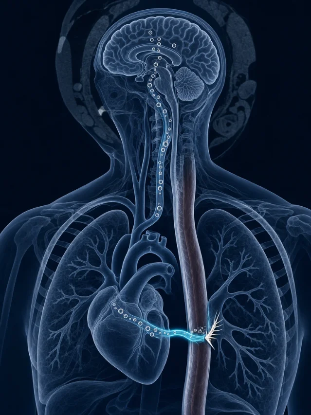
CT of the chest confirmed the impacted bone and visible air bubbles in the pulmonary vein itself. CT of the head showed punctate hypodense foci — air bubbles — in the right frontal white matter, and CT perfusion confirmed decreased cerebral blood flow in the affected zone. MRI on day six confirmed cortical infarctions in the right hemisphere. 1
The bone was removed by urgent endoscopy under general anaesthesia, with mucosal clipping for haemostasis at the perforation site. The authors (Kawai R et al.) described the case as underscoring the diagnostic importance of multimodal imaging and procedural planning for the management of cerebral air embolism caused by oesophageal perforation.
The vertebral route — bone piercing the oesophageal wall into a pulmonary vein — requires a highly specific anatomical alignment that most ingested foreign bodies never achieve. This case is published in BMJ Case Reports (DOI: 10.1136/bcr-2026-272690).
The standard clinical teaching is that air embolism via the venous system goes to the right heart and causes pulmonary complications. Left-sided air embolism, the kind that reaches the brain, is far less commonly considered in a patient who collapsed the day after eating dinner. The fish bone was the last thing on anybody's differential.
The irrigant that stopped a man's heart
A man in his 20s was undergoing wound debridement and external fixation for a femoral fracture from a blast injury — the kind of messy, contaminated trauma wound where irrigation with an antiseptic is a standard intraoperative step. Anaesthesia had been induced with propofol and nalbuphine; his airway was secured with an i-gel laryngeal mask; everything was proceeding normally. 2
About 45 minutes in, the surgical team irrigated the semi-closed wound with hydrogen peroxide. Immediately after: a sudden fall in end-tidal CO₂, then tachycardia, then rapid progression to asystole. The heart stopped.
CPR started immediately. Tracheal intubation was performed during the first compression cycle. Return of spontaneous circulation came six minutes later. The patient recovered fully — no neurological deficit, no cardiovascular sequelae.
The mechanism: hydrogen peroxide in a confined cavity does not simply clean the wound and rinse away. It liberates oxygen gas. In a wound cavity with compromised vascularity from the blast injury, that oxygen entered the venous system and caused a massive oxygen embolism to the right heart — producing the same catastrophic haemodynamic collapse as an air embolism, but from a source that every trauma surgeon has handled hundreds of times. 2
The authors (Sharafat MA et al.) wrote: "This case highlights the need to limit or avoid the use of hydrogen peroxide as irrigant whenever possible, and exercise caution and preparedness whenever its use becomes unavoidable." 2
Hydrogen peroxide has been documented as a cause of gas embolism in isolated prior reports. Penetrating and blast injuries, with their disrupted tissue planes and venous channels, are presumably higher-risk than clean surgical wounds — but the case adds to a pattern suggesting the risk is underappreciated in intraoperative practice. The case was published in BMJ Case Reports on June 3, 2026 (DOI: 10.1136/bcr-2025-271477).
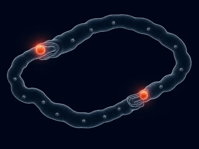
Thirteen tumors, eleven invisible, and a patient who said no
A woman in her early 50s arrived at hospital with acute abdominal pain. Emergency laparoscopy, converted to open laparotomy, found two synchronous jejunal intussusceptions — sections of small bowel telescoping into themselves, the kind of obstruction that in adults almost always indicates a structural lead point. 3
The surgeons, once inside, kept counting. The final tally was 13 separate tumors throughout the jejunum. Only two of them — the ones acting as lead points — had caused symptoms. The other eleven were clinically silent: no pain, no obstruction, no sign on imaging that anything was wrong with them. All 13 were resected. Pathology confirmed multifocal histiocytic sarcoma, an exceedingly rare malignancy of the small intestine, with mesenteric nodal metastasis. 3
Histiocytic sarcoma (HS) is a malignancy of mature histiocytes — cells normally involved in tissue repair and immune function. Primary HS of the small intestine is among the rarest of rare GI malignancies; the multifocal variant, with 13 independent tumors, is not something the surgical literature prepared anyone for.
The patient was recommended adjuvant chemotherapy. She declined treatment. She died 12 months after surgery. 3
The authors (He B, Liu M, and Cheng S-B) noted this case underscores the need to consider rare neoplasms in adult intussusception, perform thorough intraoperative exploration, and obtain extensive histopathology and immunohistochemistry for diagnosis. The case was published June 5, 2026 (DOI: 10.1136/bcr-2026-272247).
Adult intussusception differs from the pediatric form, where the bowel folds on itself without a structural cause. In adults, something is nearly always responsible — and that something, as this case shows, can be present in a quantity that only the surgeon's eye inside the abdomen can find.
The skin that announced cancer, then took it back
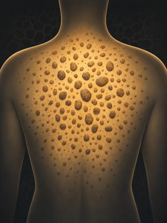
An 80-year-old man with an underlying malignancy presented with a rash of four months' standing. Physical examination found three simultaneous skin findings: exfoliative erythroderma (widespread red, scaling skin over a large body surface area), palmoplantar keratoderma (pathologically thickened skin on the palms and soles), and numerous brown, raised papules scattered across his back. 4
The patient reported that the brown papules — seborrheic keratoses (the kind of benign, warty growth that many people accumulate slowly over decades) — had appeared suddenly and concurrently with the other skin changes. This is the Leser-Trélat sign: the abrupt, simultaneous eruption of multiple seborrheic keratoses as a paraneoplastic phenomenon, signaling the presence of an internal malignancy.
The Leser-Trélat sign is listed in dermatology textbooks, but its existence as a reliable paraneoplastic marker has been debated in the literature — in part because seborrheic keratoses are so common in older adults that coincidental appearance alongside a cancer diagnosis is statistically plausible. What makes this case notable, according to the title, is the dramatic nature of both onset and resolution: the keratoses appeared suddenly with the cancer diagnosis and resolved when the underlying malignancy was treated. 4
That resolution pattern is the clinical argument for a real paraneoplastic mechanism rather than coincidence. Growth factors and cytokines secreted by the tumor (including epidermal growth factor, TGF-α, and others) are the proposed mechanism for driving the sudden keratosis proliferation — remove the tumor source, and the signal stops. The case was published June 1, 2026 in BMJ Case Reports (DOI: 10.1136/bcr-2025-270056).
The cancer drug that made the thyroid stage a coup
An 80-year-old man with advanced lung cancer was receiving nivolumab and ipilimumab — a dual immune checkpoint inhibitor (ICI) regimen that blocks PD-1 and CTLA-4, respectively, removing two of the brakes the immune system normally keeps engaged. He was hospitalized for multiple rib fractures when he developed fever, delirium, atrial fibrillation, and marked thyrotoxicosis. The clinical picture met criteria for thyroid storm — a life-threatening hypermetabolic crisis carrying substantial mortality risk even when promptly treated. 5
ICI-associated thyroid dysfunction is a well-recognized immune-related adverse event. The usual mechanism is destructive thyroiditis: the unshackled immune system attacks the thyroid follicles, causing them to rupture and release all stored thyroid hormone at once — a transient thyrotoxicosis that tends to be self-limiting. Management is largely supportive: beta-blockers for symptoms, monitoring for the hypothyroidism that typically follows.
What the authors (Hagiwara J et al.) found in this case was different. Thyroid-stimulating immunoglobulins (TSI) were strongly positive — confirming that the mechanism was not destruction of the gland but active Graves' disease: the immune system was generating antibodies that mimic TSH and drive the thyroid into sustained, progressive overdrive. The gland was not releasing stored hormone; it was being told to produce more. 5
The distinction has direct management consequences. Destructive thyroiditis does not respond to antithyroid drugs (thionamides like methimazole), because the thyrotoxicosis is from release of stored hormone rather than active synthesis. Graves' disease does respond to thionamides and may require radioactive iodine or surgery in refractory cases. Treating ICI-Graves' as if it were ICI-thyroiditis would leave the underlying driver unaddressed.
As Hagiwara et al. wrote: "This case highlights that ICI therapy-associated thyroid storm can arise not only from destructive thyroiditis but also from Graves' disease." 5
ICI-related endocrinopathy has generated a growing body of literature as checkpoint inhibitor use has expanded; within that body, the sub-categorization of "which thyroid mechanism" matters practically because treatment diverges at precisely the point where a patient is in crisis. This case published June 1, 2026 (DOI: 10.1136/bcr-2026-272027) adds documented evidence that ICI-triggered thyroid storm can arise via Graves' disease — not only via destructive thyroiditis.
Four autoimmune diseases, one viral trigger, one breast ulcer
A woman in her early 70s was already managing two diagnosed autoimmune conditions when this story started: rheumatoid arthritis (RA), a chronic joint-destroying inflammatory disease, and giant cell arteritis (GCA), an autoimmune vasculitis of large vessels that causes headache, jaw pain, and can cause permanent vision loss. She had also had MRI-confirmed bilateral optic neuritis — inflammation of the optic nerves, a third autoimmune condition — and had been placed on tapering systemic corticosteroids for GCA management. 6
Then she developed shingles — herpes zoster reactivation, which occurs when the varicella-zoster virus, dormant in the dorsal root ganglia after a childhood chickenpox infection, reactivates under conditions of immune stress. Corticosteroids are a known predisposing factor.
Shortly after the shingles, she developed painful ulcers on her breast. The morphology was distinctive: the ulcers had the violaceous (blue-purple) undermined edges and rapid tissue destruction that characterize pyoderma gangrenosum (PG) — a severe autoinflammatory neutrophilic dermatosis that is not an infection despite its name. The ulcers met Delphi diagnostic consensus criteria for PG. 6
The connection between the shingles and the PG outbreak is pathergy — a phenomenon in which minor trauma or infection triggers PG at or near the affected site. Pathergy is an established feature of PG, but it is more commonly triggered by surgical wounds, intravenous line insertion, or minor skin trauma than by viral infections. The herpes zoster here served as a systemic immune provocation that, in a patient already running multiple autoimmune processes simultaneously, tipped the balance into a fourth.
The fourth condition brought the total to: GCA, bilateral optic neuritis, rheumatoid arthritis, and now pyoderma gangrenosum — all active simultaneously in one patient. Intralesional corticosteroid injections, delivered directly into the ulcer margins, achieved complete healing within one month. This worked despite the fact that ongoing low-dose systemic steroids — already being administered for GCA — had not prevented the PG outbreak. 6
The case, published June 2, 2026 in BMJ Case Reports (DOI: 10.1136/bcr-2025-271851), is a reminder that PG's most dangerous feature is not its appearance but the risk of making it worse. PG lesions are famously susceptible to pathergy — debridement, biopsy, and other interventions that would be standard for an infected wound can accelerate tissue destruction. The correct diagnosis redirected this patient from what might have been an escalating surgical spiral toward a treatment that closed the wound in four weeks.
Cover image: AI-generated editorial composite, created for this article.
参考来源
- 1Cerebral air embolism from oesophageal-pulmonary vein fistula induced by fish bone ingestion — BMJ Case Reports
- 2Suspected venous oxygen embolism following hydrogen peroxide wound irrigation causing intraoperative asystole — BMJ Case Reports
- 3Multifocal jejunal histiocytic sarcoma presenting with dual intussusception — BMJ Case Reports
- 4Dramatic onset and resolution of Leser-Trélat sign — BMJ Case Reports
- 5Thyroid storm triggered by immune checkpoint inhibitor therapy — BMJ Case Reports
- 6Pyoderma gangrenosum as a manifestation of multisystem autoimmunity in a patient with giant cell arteritis, optic neuritis and rheumatoid arthritis — BMJ Case Reports
围绕这条内容继续补充观点或上下文。